Los Angeles Orthopedic

Cartilage Reconstruction for Joint Damage Relief

When trauma or early arthritis damages the cartilage in your knee, hip, or ankle, joint-preserving cartilage reconstruction can rebuild the surface — keeping your natural joint and delaying replacement. LAOSS surgeons offer the full spectrum, from microfracture to autologous cell-based MACI.

Cartilage Reconstruction at LAOSS orthopedic clinic in Los Angeles — board-certified specialists, same-day appointments
Live · Now Accepting

Experts in cartilage reconstruction care.

Surgical and non-surgical options at LAOSS.

15+
Years caring
Same-day appointments
Often available
★★★★★
4.9 · 7,500+ reviews

Common cartilage reconstruction concerns we treat

  • Pain that limits walking, lifting, or sleep
  • Stiffness, swelling, or reduced range of motion
  • Sports injuries — acute or overuse
  • Arthritis or post-traumatic joint changes
  • Conditions other doctors couldn't resolve

What sets LAOSS apart

  • Same- or next-day appointments at eight Los Angeles–area offices
  • On-site imaging; PT coordinated with your in-network provider
  • Conservative-first care, surgery only when needed
  • Board-certified specialists, not generalists
Key takeaways
  • Cartilage reconstruction is joint-preserving surgery — microfracture, MACI, OATS, and osteochondral allograft — that rebuilds the cartilage surface instead of replacing the whole joint.
  • Best suited for younger, active patients (typically 20–50) with focal cartilage damage from sports injury, OCD, or post-traumatic lesions — not widespread arthritis.
  • Most procedures are done arthroscopically. The right technique depends on lesion size, depth, location, and your activity goals.
  • Recovery runs 4–9 months; return to running is usually around month 4, contact sports around 9–12. Same-day evaluation available at all eight LAOSS offices.
Overview

What is cartilage reconstruction?

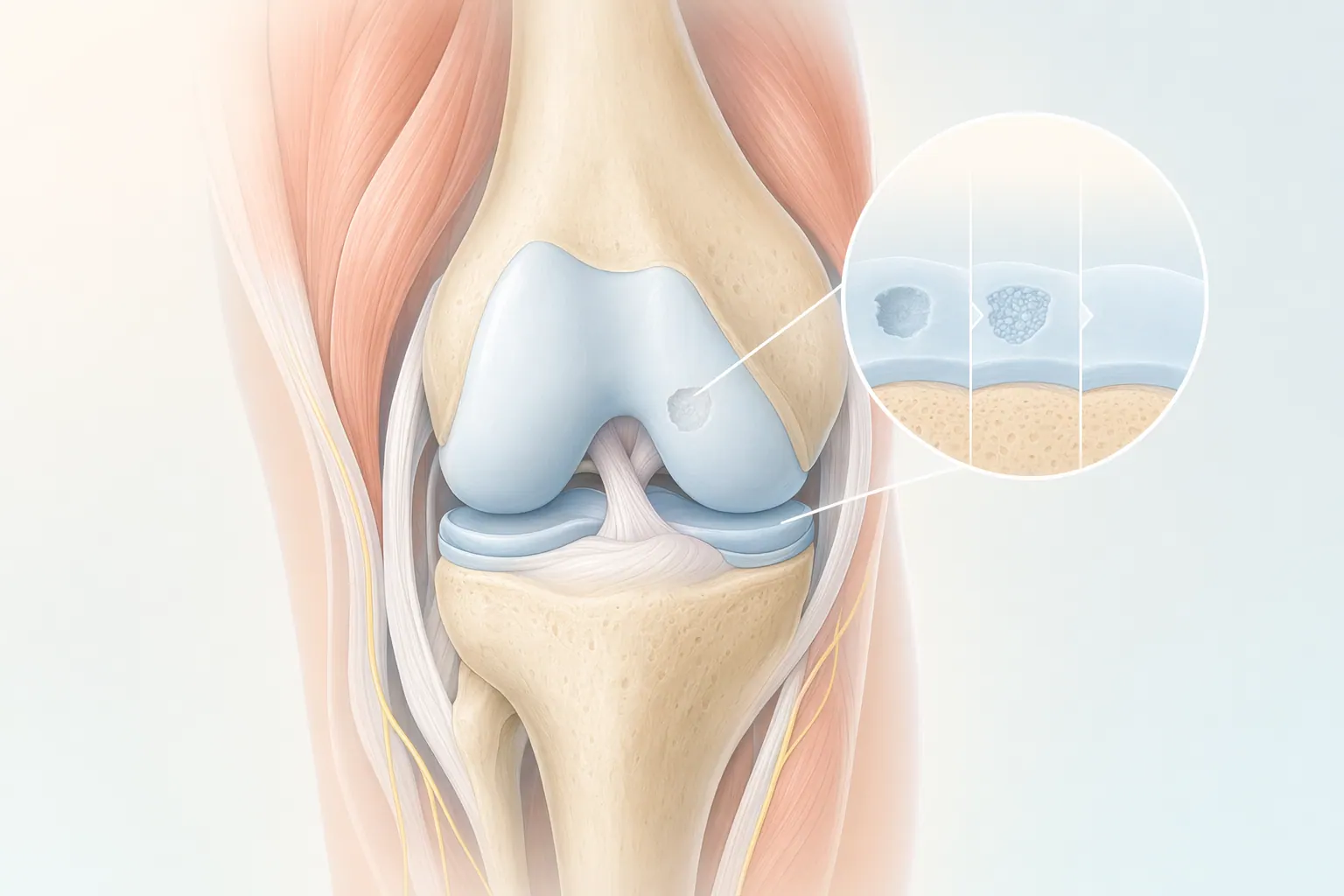
Cartilage reconstruction is a category of joint-preserving surgery that rebuilds damaged cartilage instead of replacing the whole joint. When the smooth articular surface inside a joint is lost — from a sports injury, a fall, osteochondritis dissecans (OCD), or an early-stage focal arthritis lesion — LAOSS surgeons can often restore that surface and keep your own joint working.

The four techniques we use most are microfracture (drilling tiny holes to recruit healing cells), MACI (Matrix-Induced Autologous Chondrocyte Implantation — your own cartilage cells grown in a lab and re-implanted), OATS (Osteochondral Autograft Transfer — moving a small plug of healthy cartilage-and-bone from a non-weight-bearing area), and osteochondral allograft (a matched donor graft for larger defects). Most are performed in the knee, but the same principles apply to the ankle, shoulder, and hip.

This is a different conversation than knee replacement. Cartilage reconstruction is for focal damage in a joint that's otherwise healthy — typically active patients in their 20s, 30s, and 40s who want to stay running, hiking, lifting, or playing for another two or three decades before a replacement is ever on the table.

Patient education

Watch: Autologous Chondrocyte Implantation (ACI)

This repairs your knee's damaged articular cartilage. That's the cartilage covering and protecting the ends of your bones. Repairing it helps you stay active. This surgery is done with two procedures. They are performed weeks apart.

Animations licensed from ViewMedica · Swarm Interactive

Illustration of cartilage reconstruction showing damaged cartilage being repaired or replaced
Cartilage reconstruction — damaged cartilage is repaired, regrown, or replaced with a graft.
Anatomy

Inside the cartilage.

Articular cartilage is the slick white surface that lets bone glide on bone. It has no blood supply of its own, which is why it heals poorly when injured. Reconstruction techniques work around that limitation — by recruiting marrow cells (microfracture), transplanting healthy tissue (OATS, allograft), or growing your own cartilage cells outside the body and putting them back (MACI). The right choice depends on lesion size, depth, location, and your activity level.

Self-orient

Common reasons we do cartilage reconstruction.

Symptoms

Common symptoms

  • Focal articular cartilage defect on MRI
  • Osteochondritis dissecans (OCD) lesion
  • Traumatic chondral or osteochondral injury
  • Early focal arthritis in a young active patient
  • Loose body or unstable cartilage flap in the joint
  • Chondromalacia patella that has progressed to full-thickness damage
  • Failed prior cartilage procedure
  • Mechanical symptoms — catching, locking, or giving way
Causes

Common causes

  • Acute sports injuries — pivoting, landing, or direct impact to the joint
  • Untreated ACL or meniscus tear that overloaded the cartilage
  • Osteochondritis dissecans, often in adolescence or early adulthood
  • Repetitive high-impact loading from running, jumping, or contact sports
  • Conservative care (PT, injections, activity modification) that didn't resolve symptoms
Diagnostics

How We Diagnose Cartilage Damage

Cartilage injuries are hard to read from the outside. The joint may swell, catch, or hurt after activity, but the damage itself is invisible without the right imaging — which is why an accurate diagnosis is the gating step before any reconstruction decision.

At LAOSS your first visit includes a focused history (mechanism of injury, prior procedures, activity goals), a hands-on joint exam, and same-day X-ray to rule out bony causes. When a cartilage lesion is suspected, we order an MRI with cartilage-specific sequences — this is what tells us the size, depth, and exact location of the defect, and whether the underlying bone is involved.

Most patients leave the first visit with a diagnosis, a candid read on whether they're a surgical candidate, and a plan — not just another referral. Set up your evaluation to get a clear answer about your joint.

Treatment options

Cartilage Procedures Performed by Our Surgeons

Not every cartilage injury needs surgery. Many patients do well with conservative care, and even when reconstruction is the right call, the specific technique depends on the size and location of the defect. Here's the full menu — and the criteria we use to choose between them.

Conservative care
Step 1

Conservative care first

For smaller lesions, recent-onset symptoms, or patients who aren't yet surgical candidates, we start here.

  • Activity modification (offloading the joint while it calms down)
  • Physical therapy — quadriceps and hip strengthening, neuromuscular control
  • Anti-inflammatory medication and a structured icing protocol
  • Corticosteroid injection for short-term symptom relief
  • Viscosupplementation (hyaluronic acid gel injection)
  • PRP (platelet-rich plasma) injection for biologic support
  • Unloader bracing for medial- or lateral-compartment damage
Surgical care
When needed

Surgical reconstruction techniques

When conservative care plateaus and the MRI shows a clear focal defect, we match the technique to the lesion.

  • Microfracture — small (<2 cm²) defects, lower-demand patients, day-surgery option
  • MACI (Matrix-Induced Autologous Chondrocyte Implantation) — medium-to-large defects (2–10 cm²), two-stage procedure, durable for active patients
  • OATS (Osteochondral Autograft Transfer) — small-to-medium defects with bone involvement, single-stage, uses your own tissue
  • Osteochondral allograft — large defects (>2.5 cm²), salvage cases, donor graft
  • Concurrent procedures — meniscus repair, ligament reconstruction, or osteotomy to correct alignment and protect the new cartilage
Surgeon expertise

Why experience matters.

Why experience matters

Cartilage reconstruction is one of the most technique-dependent procedures in orthopedics. The surgeon's case volume, lesion-matching judgment, and arthroscopic skill drive the durability of the repair.

  • Lesion-by-lesion matching of technique to defect size, depth, and location
  • Arthroscopic precision honed across hundreds of cartilage cases
  • Concurrent management of meniscus, ligament, and alignment issues
  • MRI interpretation and surgical planning done by your surgeon, not handed off

The LAOSS approach

Joint preservation is a long game. We start with the least-invasive option that has a real chance of working, escalate only when it doesn't, and plan every surgery around protecting your joint for the decades that come after it.

  • Same-day imaging and MRI coordination at most offices
  • PT coordinated in your insurance network from week one
  • Board-certified surgeons performing the procedures themselves
  • Direct access to your specialist between visits and through recovery
Candidacy

Am I a candidate?

Cartilage reconstruction works best for a specific profile — focal damage, healthy surrounding cartilage, an active patient who wants to keep their natural joint. If most of these match, an evaluation is the next step.

You may be

You may be a candidate if

These signs typically point toward an in-person evaluation with a cartilage specialist.

  • You're between roughly 20 and 50 and want to delay or avoid joint replacement
  • Imaging shows a focal cartilage defect rather than widespread arthritis
  • Pain, catching, or swelling limits running, sports, or work
  • Conservative care (PT, injections, activity modification) hasn't fully resolved symptoms
  • Your joint is mechanically stable or any instability is correctable in the same operation
Evaluation

What evaluation includes

Your first visit is built to give you a real answer the same day — including a candid read on whether reconstruction fits your case.

  • Detailed history — mechanism of injury, prior surgeries, activity goals
  • Hands-on exam focused on the affected joint
  • Same-day X-ray; MRI ordering with cartilage-specific protocol when needed
  • Lesion-matched plan (microfracture vs. MACI vs. OATS vs. allograft)
  • Clear timeline for recovery and return to your sport or job
ImportantSeek urgent evaluation for sudden severe joint pain after an injury, locking that won't release, rapid swelling, or any sign of infection (fever, increasing redness, or warmth at a surgical site).
Recovery

Your cartilage reconstruction recovery roadmap.

Cartilage takes time to mature. The repair is fragile in the early weeks and gets stronger month by month — which is why protected loading and patient adherence are as important as the surgery itself.

01Weeks 0–6

Protected phase

Early on we protect the repair from compressive load while keeping the joint moving. The cartilage is still healing in — pushing too hard now is the most common way to compromise the result.

  • Protected weight-bearing per procedure (often crutches for 4–6 weeks)
  • Continuous passive motion or daily ROM work to nourish the cartilage
  • Brace per surgeon protocol; ice and elevation
  • Quad activation and gentle hip strengthening from week one
02Weeks 6–16

Progressive rehabilitation

Load increases on a clear schedule. PT shifts from protection to rebuilding strength, balance, and confidence in the joint.

  • Progressive weight-bearing and gait normalization
  • Strengthening, proprioception, and neuromuscular control
  • Stationary cycling, pool work, and elliptical added in
  • Coordinated PT through your in-network provider
03Months 4–12

Return to activity

Light running typically returns around month 4; pivoting and contact sports closer to 9–12 months. We benchmark each milestone before clearing it.

  • Return-to-run progression with functional testing
  • Sport-specific drills before return-to-play clearance
  • MRI check-in at one year for larger reconstructions
  • Maintenance plan to protect the joint for the long run
Risks & considerations

What to weigh before you decide.

We talk through the risks and benefits with every patient — informed consent is a conversation, not a form.

General

General considerations

Every orthopedic operation carries a small set of standard risks. We screen, prepare, and monitor for these on every patient.

  • Infection (rare with modern technique and prophylaxis)
  • Bleeding, swelling, or bruising at the surgical site
  • Reaction to anesthesia or medications
  • Blood clot risk; managed with movement and prophylaxis as needed
Specific

Cartilage-specific considerations

Cartilage reconstruction has its own profile of risks tied to how the repair heals. These are part of every pre-op conversation.

  • Stiffness from prolonged protected weight-bearing — managed with structured PT
  • Incomplete cartilage fill or repair that doesn't fully take
  • Need for a follow-up procedure in a small percentage of cases
  • Graft-site discomfort with OATS; donor matching delay with allograft
  • Two-stage timeline with MACI (biopsy, then implantation weeks later)
Your care team

Meet the Cartilage Specialists at LAOSS

Cartilage reconstruction sits at the intersection of sports medicine and joint preservation. At LAOSS, you'll work with board-certified surgeons who do this work routinely — matching technique to lesion, coordinating PT through recovery, and planning every procedure around protecting your natural joint for the long run.

Patient reviews

What patients say about us.

★★★★★4.97,500+ Google reviews
5 stars. Got in same day for a soccer injury, X-ray right there in the office, had a plan before I left. Couldn't ask for more.
Diego Martinez
Boyle Heights, CA · 22 April 2025
Explore related care

Find care by body area.

Jump to a nearby condition page and compare treatment paths across the body.

FAQ

Common cartilage reconstruction questions

  • Knee replacement removes the joint surfaces and replaces them with metal and plastic implants — it's used for advanced, widespread arthritis. Cartilage reconstruction preserves your natural joint by rebuilding a small, defined area of damage. It's intended for younger, active patients with focal cartilage loss in an otherwise healthy joint, with the goal of delaying or avoiding replacement entirely.
  • The choice depends on the size, depth, and location of the defect, the condition of the underlying bone, and your activity goals. Smaller lesions in lower-demand patients often do well with microfracture. Medium-to-large defects in active patients are usually best served by MACI. OATS works well when bone is involved and the defect is small to medium. Larger defects or salvage cases may need an osteochondral allograft. Your LAOSS surgeon will recommend the option that fits your MRI and your life.
  • Most patients return to light running around 4 months after surgery, with progression to higher-impact activity over the months that follow. Pivoting sports and contact activity typically resume around 9–12 months. The exact timeline depends on which technique you had, how the repair matured on imaging, and how you progress through functional benchmarks in PT.
  • No surgery is a permanent guarantee, but well-selected, well-executed cartilage reconstruction can last many years — often a decade or more — and frequently delays the need for joint replacement. Durability depends on the technique used, the size of the original defect, the alignment and stability of the joint, and how well the joint is protected during recovery and afterward.
  • Most cartilage reconstruction procedures are covered when medical necessity is documented — typically a focal defect on MRI, failed conservative care, and an appropriate candidate. MACI and osteochondral allograft sometimes require prior authorization. Our team handles the paperwork and verifies your coverage before scheduling.
  • If you have persistent joint pain, swelling, catching, or giving way after an injury — especially if it limits running, sports, or work — an evaluation is the right next step. Cartilage damage doesn't heal on its own, and untreated focal lesions can grow over time, narrowing your reconstruction options later.
Ready when you are

Don't wait on pain.

Book a visit with a cartilage reconstruction specialist at any of our eight Los Angeles–area offices.

Booking now
21 specialists · 8 offices
Greater Los Angeles
On-site X-raySame visit
Most insurers acceptedIn-network
Call usBook online