Plantar fasciitis is one of the few orthopedic diagnoses we can usually make on history and exam alone — no MRI needed. When you describe stabbing heel pain on your first steps in the morning that eases as you walk, you've already told us most of what we need to know. Confirming it takes about two minutes of hands-on exam.
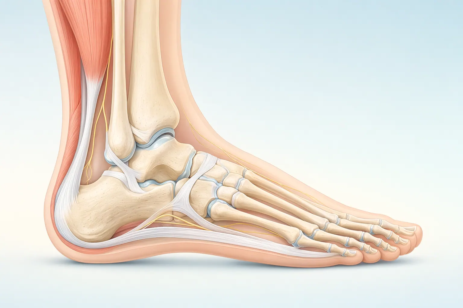
At your first visit, your LAOSS foot and ankle specialist will ask when the pain started, what your typical day looks like (how long you stand, how far you run, what shoes you live in), and what's made it better or worse. Then we examine the foot: we palpate the inside-front of the heel where the fascia attaches, test calf flexibility (a tight Achilles is almost always part of the picture), look at your arch shape, and watch you walk.
We use on-site X-ray when we need to rule out other causes — a calcaneal stress fracture, a fat pad atrophy, or in older patients, signs of inflammatory arthritis. A heel spur on X-ray, by the way, is usually not the cause of your pain. Most people with plantar fasciitis don't have one, and most people with one don't have pain. We add diagnostic ultrasound or MRI only if the picture isn't clean — for example, if we suspect a partial plantar fascia tear in a runner, or if symptoms haven't responded to 6+ months of good conservative care.
Most patients leave the first visit with a confirmed diagnosis, a written stretching protocol, footwear and orthotic guidance, and a follow-up scheduled at 4–6 weeks to measure progress.