Los Angeles Orthopedic
Foot & Ankle · Conditions A–Z

Osteochondral Lesion of the Talus cartilage and bone injury

An osteochondral lesion of the talus is an area of damaged cartilage and the bone beneath it on the dome of the talus, the ankle's central bone, often following a sprain or fracture. Same-day or next-day evaluations with on-site imaging are available across eight LA-area offices.

Los Angeles orthopedic specialist evaluating a patient for osteochondral lesion of the talus — LAOSS board-certified care across eight LA offices
Live · Now Accepting

Experts in foot & ankle care.

Surgical and non-surgical options at LAOSS.

15+
Years caring
Same-day appointments
Often available
★★★★★
4.9 · 7,500+ reviews

Common foot & ankle concerns we treat

  • Pain that limits walking, standing, or sleep
  • Stiffness, swelling, or reduced range of motion
  • Sports injuries — acute or overuse
  • Arthritis or post-traumatic joint changes
  • Conditions other doctors couldn’t resolve

What sets LAOSS apart

  • Same- or next-day appointments at eight Los Angeles–area offices
  • On-site imaging; PT coordinated with your in-network provider
  • Conservative-first care, surgery only when needed
  • Board-certified specialists, not generalists
Key takeaways
  • An osteochondral lesion of the talus (OLT) is a cartilage-and-bone injury on the talar dome, frequently caused by an ankle sprain or fracture that didn't fully heal.
  • Deep, aching ankle pain with activity, catching, swelling, and a sense of giving way are the most common signs.
  • Stable, small lesions often respond to a period of protected weight-bearing, bracing, and physical therapy; an MRI confirms the size and stability.
  • When pain persists or the cartilage fragment is unstable, ankle arthroscopy and cartilage-restoration procedures are highly effective options.
Overview

What is osteochondral lesion of the talus?

An osteochondral lesion of the talus, often called an OLT or talar dome lesion, is an injury to the smooth cartilage cap on the top of the talus and the bone directly beneath it. Because the talus sits at the center of the ankle joint, even a small lesion can produce deep, nagging pain that doesn't behave like an ordinary sprain. Many OLTs are first noticed when ankle pain lingers for weeks or months after an injury that was expected to heal.

Most OLTs are discovered after an ankle sprain or fracture, and a fair number are missed on initial X-rays because cartilage and early bone changes don't always show up. That's why an MRI is so useful here — it shows the size of the lesion, whether the underlying bone is bruised or cystic, and whether the cartilage fragment is stable or loose.

Treatment is conservative-first for stable lesions, and surgical when the cartilage is unstable or symptoms persist. Below, we walk through the symptoms and causes we see most often, how we confirm the diagnosis, and the full range of non-surgical and surgical options — explained in plain English.

Patient education

Watch: Osteochondral Lesion of the Talus

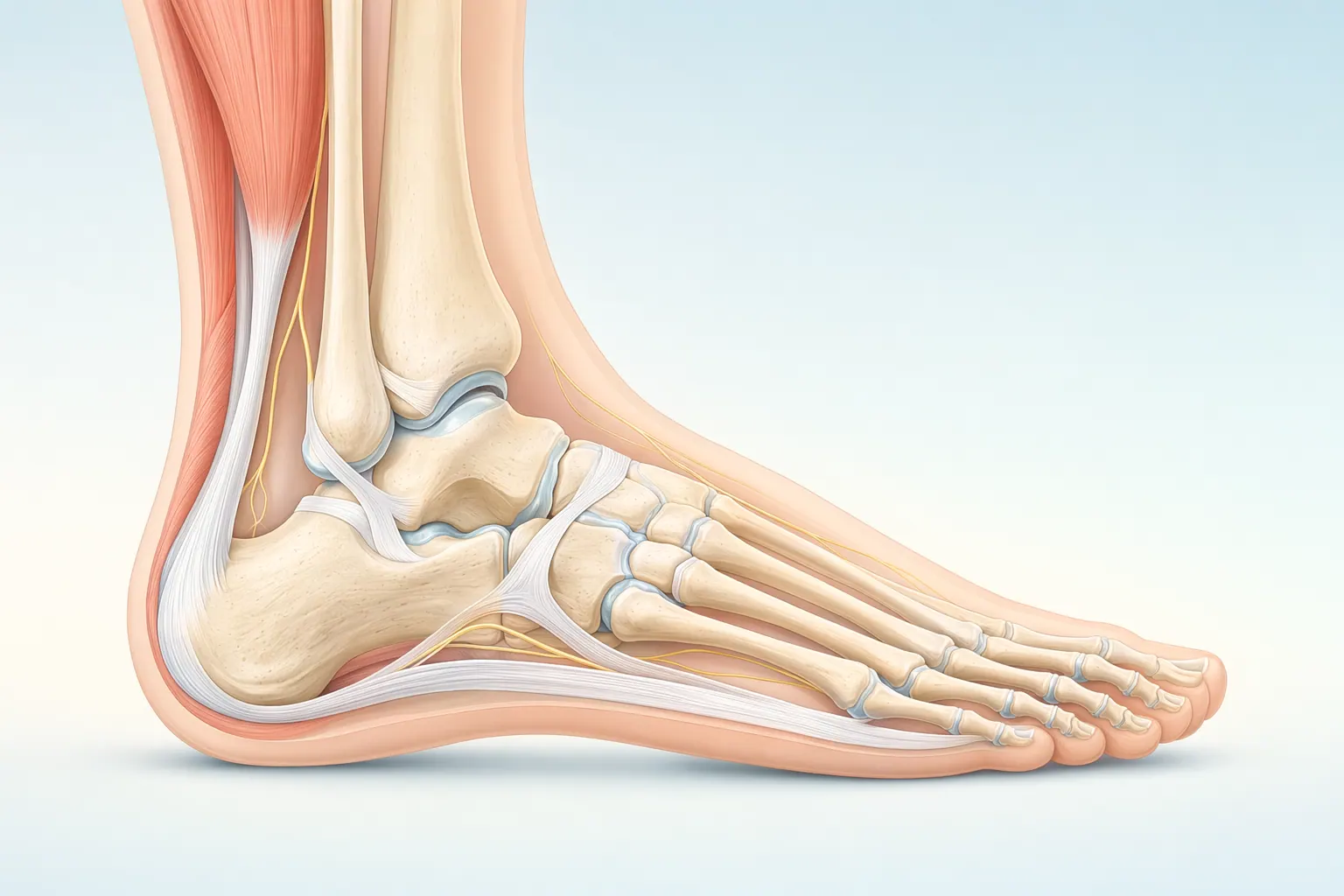
This is an injury of the protective cartilage on the top of the talus (the ankle bone). You'll find this cartilage where the talus touches the tibia and fibula (the bones of the lower leg). An osteochondral lesion can be a painful problem.

Animations licensed from ViewMedica · Swarm Interactive

Anatomical illustration of the foot and ankle showing the tibia, talus, calcaneus, and plantar fascia
Anatomy of the foot & ankle — tibia, talus, calcaneus, metatarsals, and the plantar fascia.
Anatomy

Inside the foot & ankle.

The foot and ankle have 26 bones, more than 30 joints, and over 100 ligaments and tendons. The plantar fascia spans the bottom of the foot, the Achilles tendon anchors the calf to the heel, and the ankle is a hinge that handles every step you take. Most foot and ankle problems trace back to overload, alignment, or footwear that doesn’t match the way your foot is built.

Self-orient

When osteochondral lesion of the talus shows up.

Symptoms

Common symptoms

  • Deep, aching ankle pain that worsens with weight-bearing and activity
  • Persistent ankle pain that lingers weeks or months after a sprain
  • Swelling that comes and goes, especially after exercise
  • Catching, clicking, or locking inside the ankle joint
  • A sense of the ankle giving way or feeling unstable
  • Stiffness and reduced range of motion in the ankle
Causes

Common causes

  • A prior ankle sprain, particularly a rolling (inversion) injury
  • An ankle fracture that involved the joint surface
  • Repetitive impact or twisting from sports and running
  • Disrupted blood supply to a small area of the talar bone
  • Chronic ankle instability that repeatedly loads the talar dome
Diagnostics

How we diagnose osteochondral lesion of the talus

You want answers, fast — and we’re built to give them. Most patients leave their first LAOSS visit with a clear diagnosis and a written plan, not another referral chain.

Here’s what your initial visit for osteochondral lesion of the talus typically looks like:

  • Detailed history — when it started, what makes it better or worse, what you’ve already tried
  • Focused exam of the foot & ankle — range of motion, stability, strength, specific provocation tests
  • On-site imaging at most offices: X-ray for bone, ultrasound or MRI when soft-tissue detail is needed
  • A written plan with options ranging from conservative care to surgical procedures, in plain English

Schedule your evaluation with a trusted Greater Los Angeles orthopedic expert today.

Treatment options

How we treat osteochondral lesion of the talus at LAOSS

Once we’ve confirmed the diagnosis, the next step is matching the right treatment to your situation. We start with the least-invasive option that fits — and escalate only when it doesn’t.

Conservative care
Step 1

Conservative care first

Non-surgical options designed to relieve pain, restore movement, and avoid the OR when possible.

  • A period of protected weight-bearing with a boot or cast to offload the lesion
  • Activity modification to reduce impact while the bone settles
  • Bracing or supportive footwear, especially with ankle instability
  • Physical therapy to restore motion, strength, and balance
  • Anti-inflammatory medication for pain and swelling
  • Repeat MRI to monitor stable lesions over time
Surgical care
When needed

Surgical options when needed

Procedures performed by board-certified foot & ankle surgeons when conservative care isn’t enough.

  • Ankle arthroscopy to inspect, debride, and remove loose cartilage fragments
  • Microfracture (marrow stimulation) to recruit healing cells into small lesions
  • Bone grafting to fill an underlying cyst or bone defect
  • Osteochondral autograft transfer (OATS) for larger lesions
  • Cartilage restoration techniques for select cases
  • Fixation of a sizable, intact cartilage fragment when it can be preserved
Surgeon expertise

Why experience matters.

Why experience matters

Foot & Ankle care is highly technique-dependent. Volume, training, and judgment together determine the outcome you actually feel six months later.

  • Precise diagnosis from imaging and exam
  • Conservative-first care that avoids unnecessary surgery
  • Surgical technique refined over thousands of cases
  • On-site imaging + coordinated PT through your in-network provider

The LAOSS approach

Our foot & ankle specialists move stepwise — start with the least-invasive option that fits your situation, escalate only when it doesn't.

  • Same-day imaging at most offices
  • PT coordinated in your insurance network
  • Board-certified surgeons performing the procedures themselves
  • Direct access to your specialist between visits
Candidacy

Am I a candidate?

If most of these match your situation, an evaluation with a foot & ankle specialist is the next step.

You may be

You may be a candidate if

These signs typically point toward an in-person evaluation with a foot & ankle specialist.

  • Ankle pain that has lingered for weeks or months after a sprain or fracture
  • Deep, aching pain inside the ankle that flares with activity or impact
  • Catching, clicking, or a sense of the ankle locking or giving way
  • Normal or unremarkable X-rays despite ongoing ankle symptoms
  • Swelling that returns each time you increase your activity level
Evaluation

What evaluation includes

Your first visit is built to give you an answer the same day, not just another referral.

  • Detailed history — onset, mechanism, what makes it better or worse
  • Hands-on exam focused on the affected joint or region
  • On-site imaging at most offices (X-ray, ultrasound)
  • Clear plan with options ranging from conservative to surgical
  • Same-day or next-day scheduling for any follow-up tests
ImportantSeek urgent evaluation for sudden severe pain, an open wound or exposed bone, foot or ankle deformity after trauma, loss of sensation, or any sign of infection (fever, spreading redness, or swelling).
Recovery

Your foot & ankle recovery roadmap.

Recovery is rarely a straight line — but a clear plan with measurable milestones makes the path predictable.

01Days 0–14

Right after care

In the first two weeks we focus on protecting the foot & ankle, calming inflammation, and restoring basic motion.

  • Activity modification with clear do/don’t guidance
  • Ice, elevation, and pain control as needed
  • Gentle range-of-motion within safe limits
  • Follow-up scheduled to track healing
02Weeks 2–8

Rehabilitation

Targeted physical therapy rebuilds strength, mobility, and confidence in the foot & ankle.

  • Progressive strengthening and neuromuscular work
  • Manual therapy and soft-tissue treatment
  • Sport- or job-specific movement re-training
  • Coordinated PT through your in-network provider
03Months 2+

Long-term care

Once function is restored, the focus shifts to keeping you there — and catching any recurrence early.

  • Return-to-activity plan with measured benchmarks
  • Home program tailored to your sport or job
  • Maintenance visits or imaging if symptoms change
  • Direct line back to your specialist if needed
Risks & considerations

What to weigh before you decide.

We talk through the risks and benefits with every patient — informed consent is a conversation, not a form.

General

General considerations

Every orthopedic intervention carries a small set of standard risks. We screen, prepare, and monitor for these on every patient.

  • Infection (rare with modern technique and prophylaxis)
  • Bleeding or bruising at the treatment site
  • Reaction to anesthesia or medications
  • Need for additional procedures in some cases
Specific

Foot & Ankle-specific considerations

Some risks are tied to the structures we're treating in the foot & ankle. We discuss these in detail at your visit so you can weigh them against the benefits.

  • Temporary stiffness or weakness during recovery
  • Incomplete pain relief in a small percentage of cases
  • Nerve or vessel irritation near the treatment area
  • Need for follow-up therapy to fully restore function
Your care team

Meet our foot & ankle doctors in the Greater Los Angeles area

At LAOSS, our foot & ankle specialists combine advanced surgical expertise with a patient-first approach. From minimally invasive arthroscopic techniques to reconstruction, fracture care, and arthritis management, our physicians bring decades of experience to every case. Trusted across Los Angeles, our team is dedicated to restoring mobility, relieving pain, and helping you return to the activities you love.

Specialists

Meet your foot & ankle specialists.

4 providers
About this care

Reliable answers for stubborn ankle pain start here

An osteochondral lesion of the talus is one of the most commonly missed causes of ankle pain that won't settle after a sprain — and at LAOSS, getting to the right diagnosis doesn't take months of referrals. With on-site imaging and same- or next-day appointments at multiple Los Angeles locations, our board-certified foot & ankle specialists can confirm whether your pain is coming from the cartilage on the talar dome and explain exactly what your MRI shows.

Whether your lesion is stable and best managed with protected weight-bearing and therapy, or it needs arthroscopic treatment, you'll get a clear, conservative-first plan and a team that performs these procedures routinely. Call or schedule online to start with a trusted foot & ankle specialist in Los Angeles.

Patient reviews

What patients say about us.

★★★★★4.97,500+ Google reviews
Wonderful staff. The MA was so kind to my elderly mom and the doctor explained everything twice so she’d remember. Felt like we were treated like family.
Patricia Aguilar
Cerritos, CA · 6 January 2025
Explore related care

Find care by body area.

Jump to a nearby condition page and compare treatment paths across the body.

FAQ

Common osteochondral lesion of the talus questions

  • We start with a focused ankle exam and history, then X-rays in-office. Because cartilage injuries often don't show on X-ray, we order an MRI to confirm the lesion's size, stability, and whether the underlying bone is involved — this drives the whole treatment plan.
  • Yes — small, stable lesions often improve with a period of protected weight-bearing in a boot, activity modification, and physical therapy. Surgery is reserved for unstable lesions or symptoms that don't settle with conservative care.
  • A lingering deep ache after a sprain is a classic sign of an osteochondral lesion of the talus, which is frequently missed on the initial visit. An MRI is the best way to confirm whether cartilage on the talar dome was injured.
  • Most cases are treated with ankle arthroscopy, where loose cartilage is cleaned out and small lesions are treated with microfracture to stimulate healing. Larger lesions may need bone grafting or a cartilage transfer, which we discuss in detail before any decision.
Ready when you are

Don’t wait on pain.

Book a visit with a foot & ankle specialist at any of our eight Los Angeles–area offices.

Booking now
21 specialists · 8 offices
Greater Los Angeles
On-site X-raySame visit
Most insurers acceptedIn-network
Call usBook online